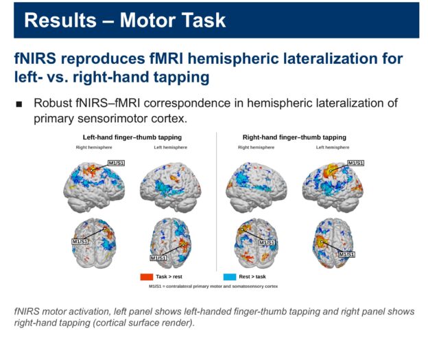
Next week I’ll be in Washington, D.C. for Neuroscience 2026, the Society for Neuroscience meeting, with a new poster from our lab: “fNIRS-fMRI Reliability for Motor and Social Perception Paradigms: Validation of fNIRS Against fMRI.” It asks a simple question that sits under almost everything we do: when fNIRS says part of the brain is active, does fMRI agree? Below is a plain-language summary and an interactive version of the poster you can click through.
The question
Our lab uses fNIRS (functional near-infrared spectroscopy), which measures hemodynamic, or blood-flow, responses with optodes placed on the scalp. Its big advantage is that it lets us study people in natural situations, including live, face-to-face interaction, which is exactly what developmental and social neuroscience needs. But fMRI is still the reference standard, and fNIRS needs to be checked against it systematically, on more than simple reference tasks. So we compared the two directly.
What we did
We used two calibrated benchmark tasks and ran each one with fNIRS (a Shimadzu LABNIRS system) and with fMRI (a Siemens MAGNETOM Prisma 3T).
- Finger-thumb tapping. People tapped with the left hand or the right hand in 15-second blocks with 15 seconds of rest, for 6 minutes, with the same timing for fNIRS and fMRI. This is a focal test: it should light up primary sensorimotor cortex on the side opposite the hand.
- Dynamic faces. People watched short videos of children’s faces smiling, moving and chewing (stimuli from Pitcher and colleagues, 2011). This targets the face network, especially the posterior superior temporal sulcus (pSTS), a region that is central to social perception.
What we found
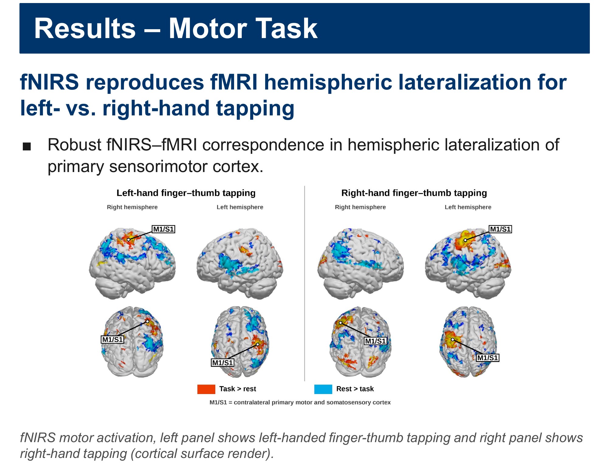
For the motor task, fNIRS reproduced the hemispheric lateralization seen with fMRI: left-hand tapping drove the right primary sensorimotor cortex and right-hand tapping drove the left, with robust correspondence between the two methods.
For the face videos, both fNIRS and fMRI picked up the dynamic-face network, including multiple sites along the pSTS. fNIRS reliably detected activity in visual and lateral-occipital cortex and in the superior and middle temporal lobes, with high spatial correspondence to the fMRI BOLD signal. Overall, the two methods showed strong spatial agreement in both tasks.
We are also working on the depth question. Continuous-wave fNIRS sees superficial cortex, roughly half the distance between a source and a detector, while fMRI is fully 3D. Using one participant’s own fMRI and anatomical scans together with a NeuroDOT sensitivity model, we are building a pipeline to estimate how well fNIRS reaches the superficial and deeper parts of the fusiform face area and the pSTS.
Why it matters
In the scanner, social stimuli have to be recorded video, and that removes two things that define a real social exchange: a recorded face can’t return your eye contact, and it can’t react to or mirror your expressions. fNIRS has neither limit, so we can study live, face-to-face interaction with real two-way gaze and facial expression. Showing that fNIRS agrees with fMRI on these benchmarks supports using it in developmental settings where fMRI is impractical and where ecological validity really matters. The honest caveat is depth: fNIRS measures cortex only to a limited depth, and any comparison with fMRI has to take that into account.
Explore the poster
Click any part of the poster to open a zoomed-in, high-resolution view of that panel with a short explanation, then use Next and Previous (or your arrow keys) to step through it in reading order. Guided tour walks you through the whole thing from the top, and you can zoom, pan or go fullscreen on the full poster. On a phone, use the list of sections under the poster. Press Esc to close a panel.
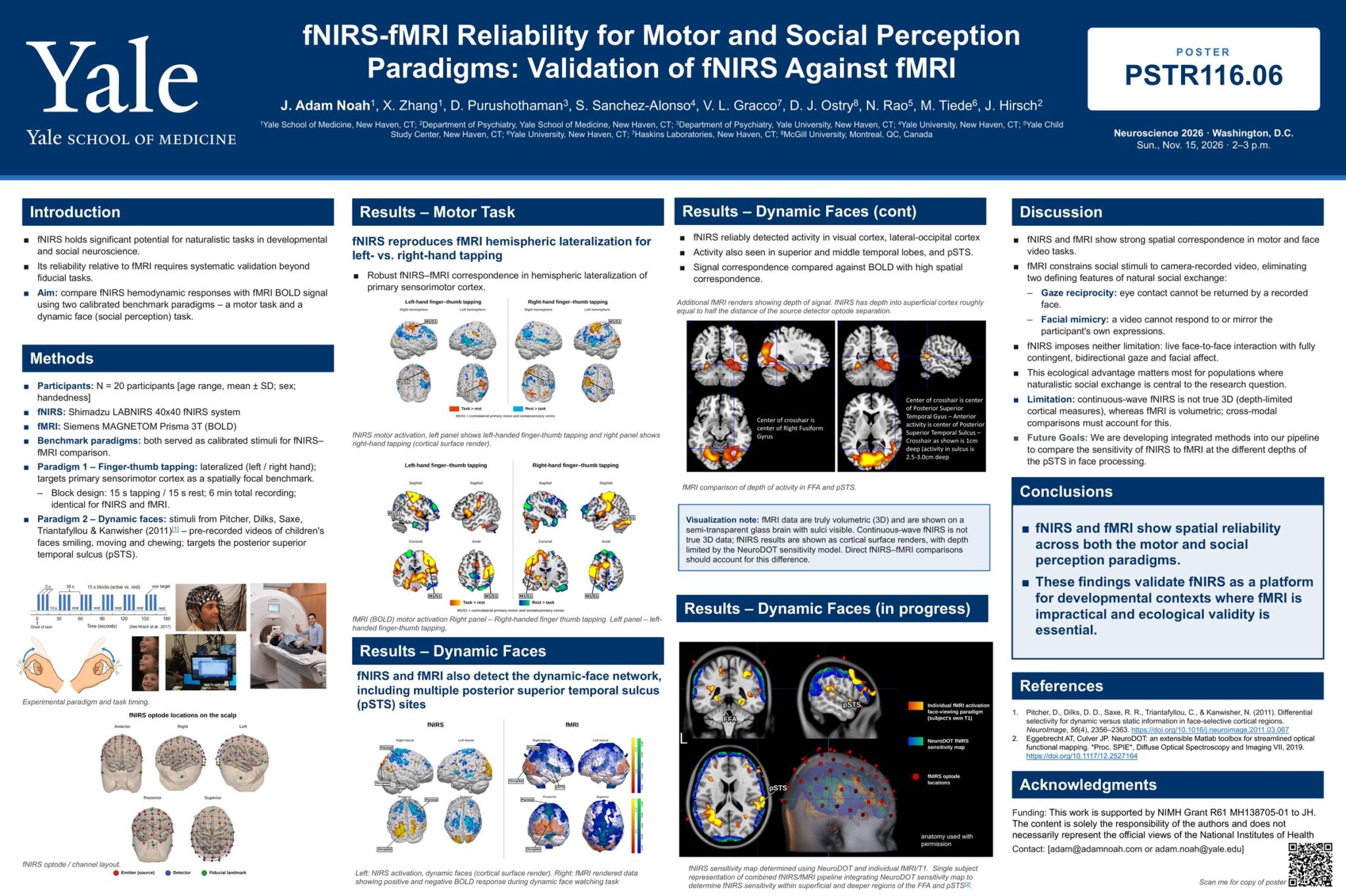
Our poster for Neuroscience 2026 in Washington, D.C.: fNIRS-fMRI Reliability for Motor and Social Perception Paradigms: Validation of fNIRS Against fMRI. It is poster PSTR116.06, listed for Sunday, Nov. 15, 2026, 2–3 p.m.
Authors: J. Adam Noah, X. Zhang, D. Purushothaman, S. Sanchez-Alonso, V. L. Gracco, D. J. Ostry, N. Rao, M. Tiede and J. Hirsch, from the Yale School of Medicine, Yale University, the Yale Child Study Center, Haskins Laboratories and McGill University.
fNIRS (functional near-infrared spectroscopy) has a lot of promise for natural, real-world tasks in developmental and social neuroscience. But before we lean on it, we need to know how well it agrees with fMRI, and that has to be tested on more than simple reference tasks.
The aim: compare fNIRS hemodynamic (blood-flow) responses with the fMRI BOLD signal using two calibrated benchmark tasks, one motor task and one dynamic-face (social perception) task.
The same benchmark tasks were run with fNIRS (a Shimadzu LABNIRS system) and with fMRI (a Siemens MAGNETOM Prisma 3T scanner).
- Task 1, finger-thumb tapping: left hand or right hand, a focal benchmark for primary sensorimotor cortex. Blocks of 15 s tapping and 15 s rest, 6 minutes in total, identical for fNIRS and fMRI.
- Task 2, dynamic faces: pre-recorded videos of children’s faces smiling, moving and chewing (stimuli from Pitcher et al., 2011), aimed at the posterior superior temporal sulcus (pSTS).
The figure shows the task timing and the recording setups.
Where the fNIRS sensors sit. The scalp models show the optode layout from the front, right, left, back and top. Red dots are emitters (light sources), blue dots are detectors, and green dots are fiducial landmarks used to line the cap up on the head.
fNIRS reproduces the left/right pattern seen in fMRI. When people tapped with the left hand, activity showed up in the right primary sensorimotor cortex (labelled M1/S1), and the reverse for the right hand.
These are fNIRS results drawn on the cortical surface: left panel for left-hand tapping, right panel for right-hand tapping. Warm colors are task > rest; blue is rest > task.
The same tapping task measured with fMRI (BOLD), shown in sagittal, coronal and axial slices. The M1/S1 labels mark primary sensorimotor cortex on the side opposite the tapping hand, which is the pattern fNIRS picked up in the previous panel. Together they show robust fNIRS–fMRI agreement in hemispheric lateralization.
Both methods pick up the face network. While people watched the dynamic face videos, fNIRS (left) and fMRI (right) both detected the dynamic-face network, including multiple sites along the posterior superior temporal sulcus (pSTS). The fMRI renders show both positive and negative BOLD responses.
fNIRS reliably detected activity in visual cortex and lateral-occipital cortex, and also in the superior and middle temporal lobes and pSTS, with high spatial correspondence to the fMRI BOLD signal.
The fMRI slices show how deep that activity sits. One crosshair marks the right fusiform gyrus; the other marks the pSTS, where the crosshair is about 1 cm deep and activity in the sulcus is 2.5–3.0 cm deep. fNIRS reaches into superficial cortex, roughly half the distance between a source and a detector optode.
A note on the pictures. fMRI data are truly 3D, so they are shown in a semi-transparent glass brain with the sulci visible. Continuous-wave fNIRS is not true 3D data, so its results are drawn on the cortical surface, with depth limited by the NeuroDOT sensitivity model. Any direct fNIRS–fMRI comparison has to account for that difference.
Work in progress: how deep can fNIRS see? For a single participant, we combined that person’s own fMRI and T1 anatomical scans with a NeuroDOT fNIRS sensitivity map. Overlaying the fMRI face activation, the fNIRS sensitivity and the optode positions lets us estimate how sensitive fNIRS is to the superficial and deeper parts of two face areas, the fusiform face area (FFA) and the pSTS.
fNIRS and fMRI line up well in space for both the motor task and the face-video task.
Why that matters: in the scanner, social stimuli have to be recorded video, which removes two defining parts of real social exchange. A recorded face can’t return your eye contact (gaze reciprocity), and it can’t respond to or mirror your expressions (facial mimicry). fNIRS has neither limit, so we can study live, face-to-face interaction with real two-way gaze and facial expression. That matters most for groups where natural social exchange is central to the question.
The limitation: continuous-wave fNIRS only measures cortex to a limited depth, while fMRI is volumetric. Next, we are building methods into our pipeline to compare fNIRS and fMRI sensitivity at different depths of the pSTS during face processing.
The take-home: fNIRS and fMRI show spatial reliability across both the motor and the social perception tasks. That supports fNIRS as a platform for developmental settings where fMRI is impractical and ecological validity is essential.
The face videos come from Pitcher, Dilks, Saxe, Triantafyllou & Kanwisher (2011, NeuroImage), and the sensitivity modelling uses the NeuroDOT toolbox (Eggebrecht & Culver, 2019).
This work is supported by NIMH Grant R61 MH138705-01 to J. Hirsch. The content is solely the responsibility of the authors and does not necessarily represent the official views of the National Institutes of Health.
Section
Download the full poster (PDF)
Come by if you’re at SfN
If you’re at Neuroscience 2026 in Washington, D.C., come find me at poster PSTR116.06 on Sunday, November 15. I’ll be at the poster from 1 p.m. (the time printed on the poster is 2–3 p.m.). Stop by and talk with me about what the lab is doing; I’d love to hear what you’re working on too.
Poster details
Title: fNIRS-fMRI Reliability for Motor and Social Perception Paradigms: Validation of fNIRS Against fMRI
Authors: J. Adam Noah, X. Zhang, D. Purushothaman, S. Sanchez-Alonso, V. L. Gracco, D. J. Ostry, N. Rao, M. Tiede, J. Hirsch
Affiliations: Yale School of Medicine; Department of Psychiatry, Yale School of Medicine; Department of Psychiatry, Yale University; Yale University; Yale Child Study Center; Haskins Laboratories; McGill University
Poster: PSTR116.06, Neuroscience 2026, Washington, D.C., Sun., Nov. 15, 2026, 2–3 p.m.
This work is supported by NIMH Grant R61 MH138705-01 to J. Hirsch. The content is solely the responsibility of the authors and does not necessarily represent the official views of the National Institutes of Health.