Our new paper is out in Frontiers in Neuroimaging, led by Deepa Purushothaman with Xian Zhang, Rahul Singh, Cenk Tek, Vinod Srihari, Joy Hirsch and me. It asks a simple question with a complicated answer: what is different in the brain when someone early in the course of psychosis looks at another person’s face, live, in the same room? The article is open access: read it here (doi:10.3389/fnimg.2026.1931240).
The problem
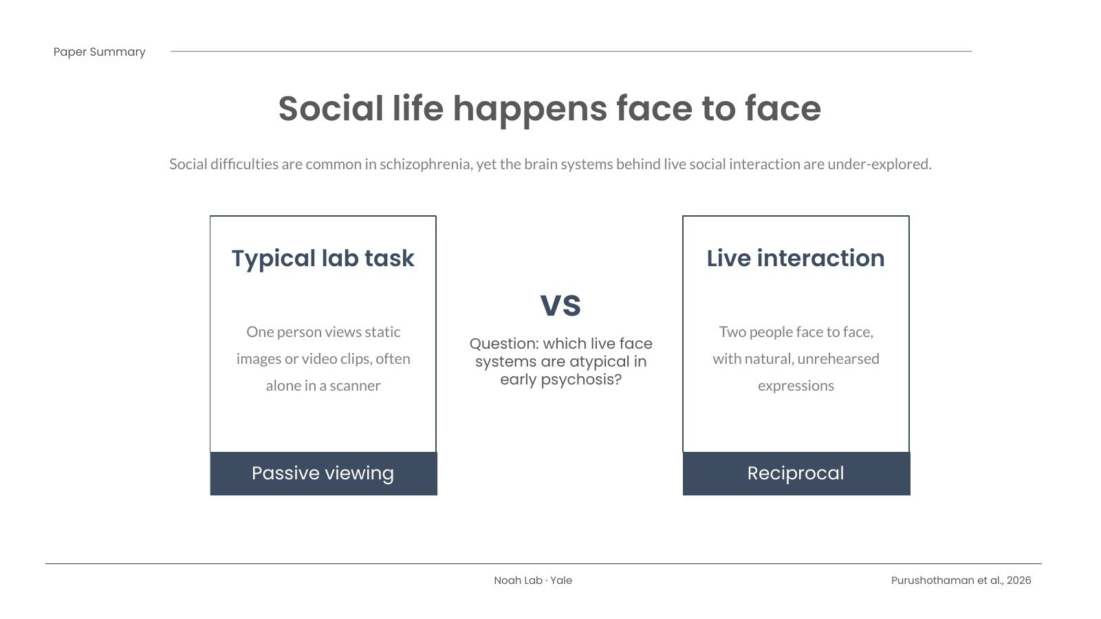
Difficulties with social interaction are common in schizophrenia, and they matter a great deal for how people do in daily life. Yet most of what we know about the social brain in schizophrenia comes from people looking at photos or video clips, usually alone, lying in a scanner. Real conversations aren’t like that. They are face to face, reciprocal and unscripted, and our earlier work suggests the brain handles a live face differently from a face on a screen.
What we did
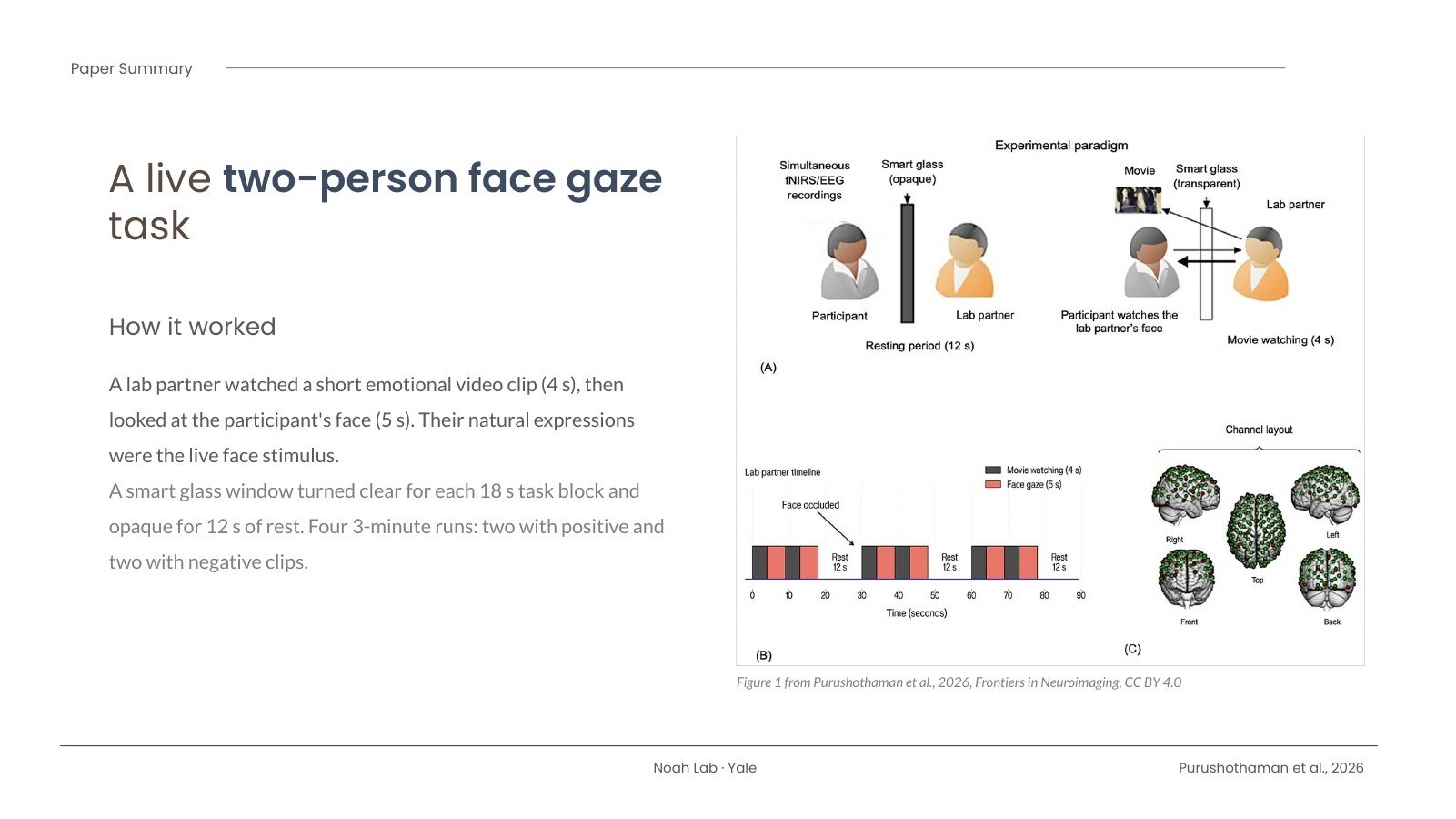
We used a two-person face gaze task that our lab has used before. A lab partner sat across a table from each participant, separated by a “smart glass” window that could switch from opaque to clear. The partner watched short, emotionally evocative video clips (cute animals, or things like spiders and decay) and then looked at the participant, so the participant saw a real person’s natural, unrehearsed expressions.
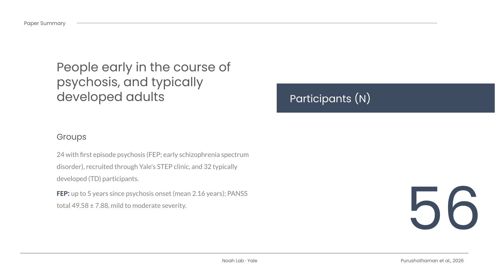
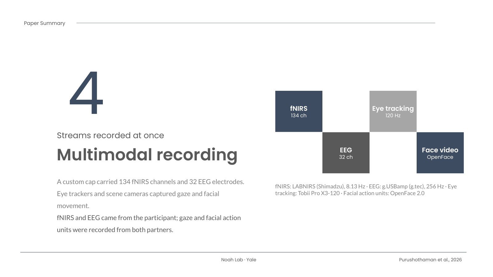
While this happened we recorded several things at once: fNIRS (134 channels) and EEG (32 electrodes) from a single cap, eye tracking, and video of facial movements analyzed with OpenFace. We compared 24 people with first episode psychosis (early schizophrenia spectrum disorders), recruited through Yale’s STEP clinic, with 32 typically developed participants.
What we found
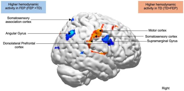
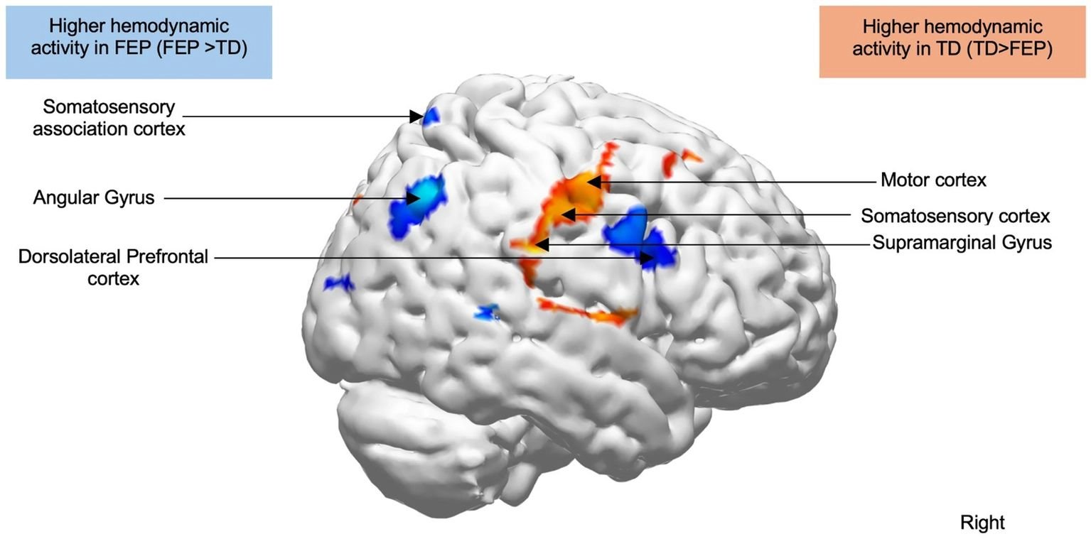
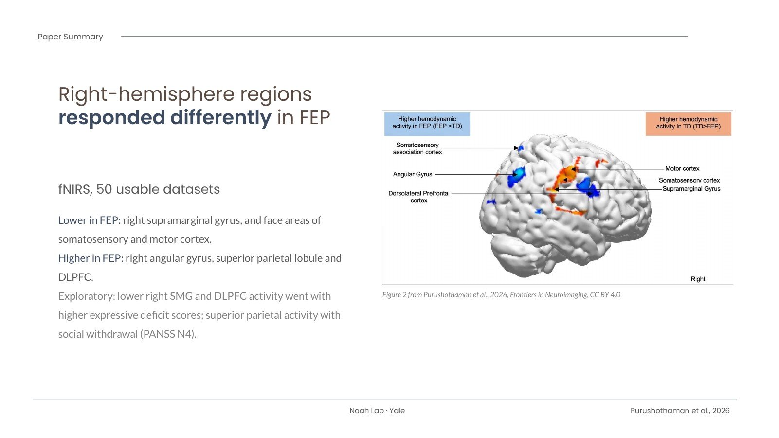
Activity in a set of right-hemisphere regions differed between the groups. The right supramarginal gyrus, a region we have repeatedly linked to live face gaze, along with the face areas of the right somatosensory and motor cortex, was less active in the first episode psychosis group. The right angular gyrus, superior parietal lobule and dorsolateral prefrontal cortex were more active.
In exploratory analyses, lower activity in the right supramarginal gyrus and DLPFC went with higher expressive deficit scores (blunted affect, poor rapport, less spontaneous conversation and slowed movement), and activity in the superior parietal region went with passive social withdrawal.
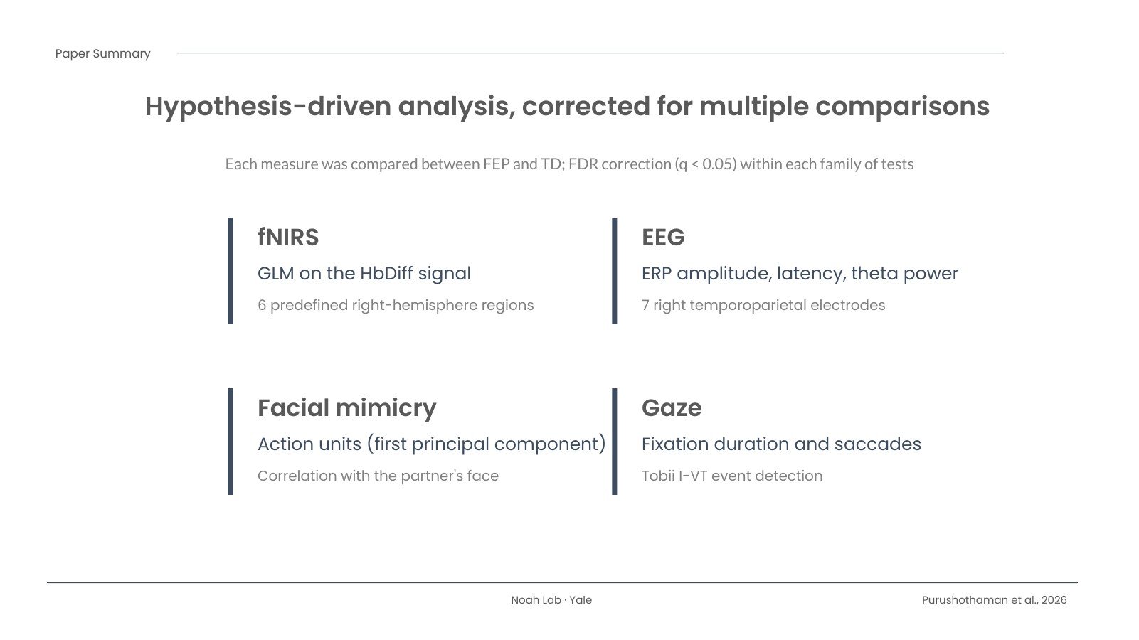
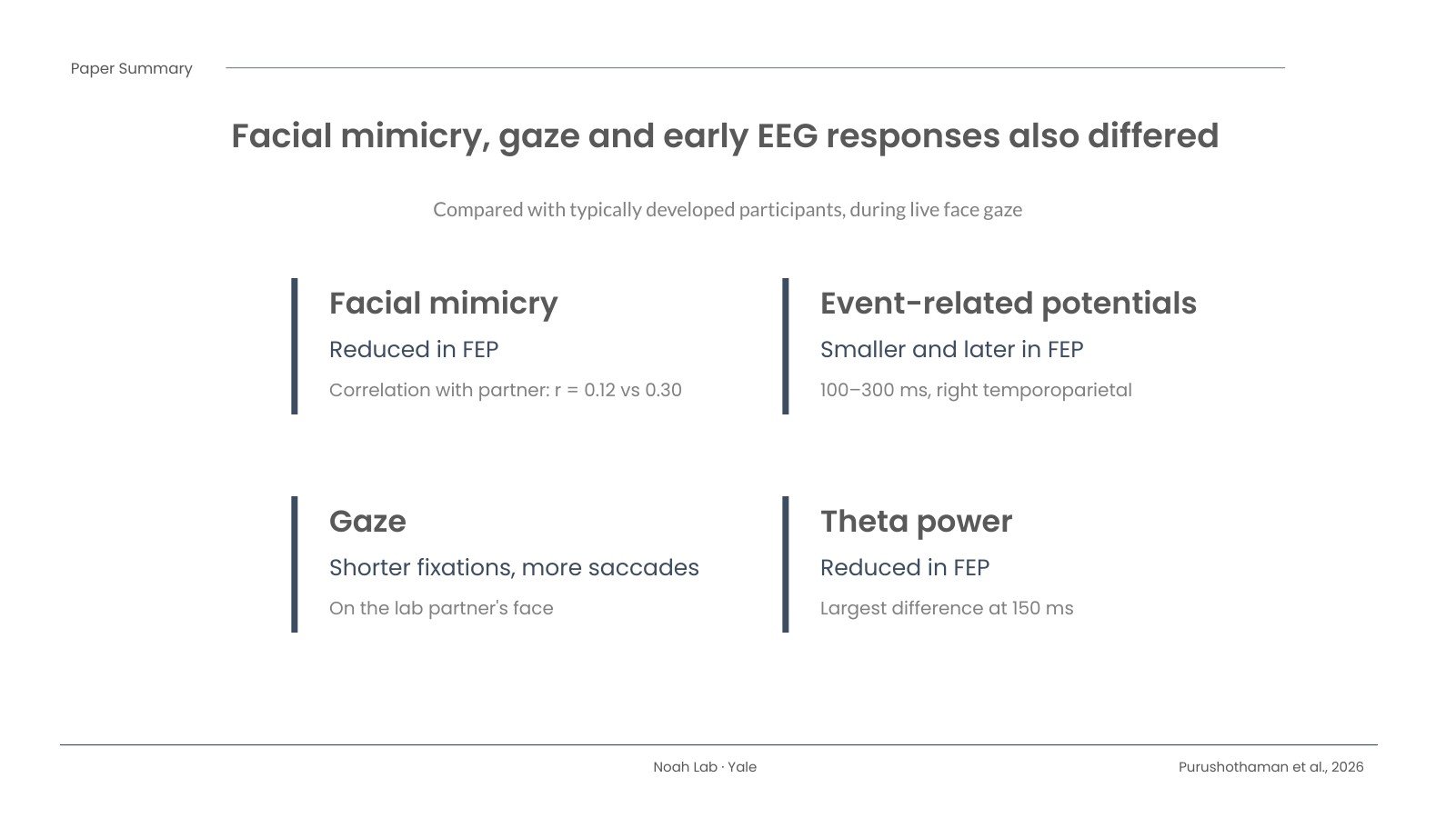
Behavior and EEG told a consistent story. Participants with first episode psychosis showed less spontaneous facial mimicry of their partner, shorter fixations on the partner’s face and more frequent saccades. In the first 100 to 300 milliseconds after the window cleared, their EEG responses were smaller over right temporoparietal electrodes and delayed over temporoparietal electrodes on both sides, and theta power was reduced. So the “where” from fNIRS and the “when” from EEG pointed to the same right temporoparietal area.
Why it matters, and what it doesn’t show
This is a step toward measuring social brain function in a setting that looks more like everyday life, at a stage of illness when people may benefit most from new kinds of support. The regions we found could become targets for future studies that test cause and effect, for example with non-invasive neuromodulation.
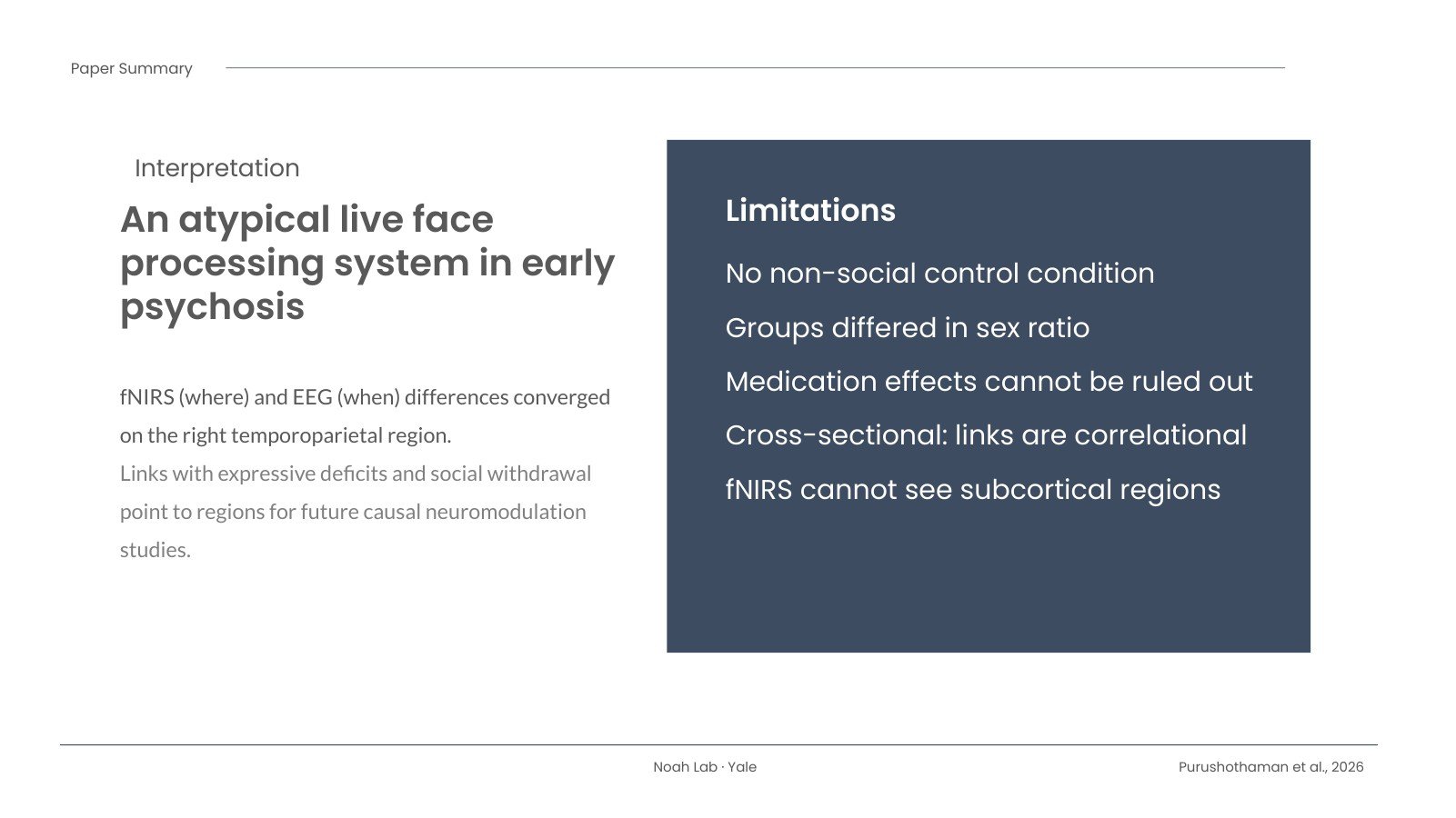
It is also important to be clear about the limits. The study is cross-sectional, so these links are correlational. There was no non-social control condition, the groups differed in sex ratio, most participants with psychosis were taking medication, and fNIRS can’t see deeper brain structures. We hope this work helps build better tools, not labels. I’m grateful to everyone who took part.
The slides
Here is a short slide summary of the paper. Use the arrows, your keyboard, or the thumbnails to move through it.
Download the slides (PowerPoint)
Read the paper
Purushothaman D, Noah JA, Zhang X, Singh R, Tek C, Srihari VH, Hirsch J. A multimodal approach to identify atypical face processing in first episode psychosis (early schizophrenia spectrum disorders). Frontiers in Neuroimaging. 2026;5:1931240. doi: 10.3389/fnimg.2026.1931240
The article is open access under a CC BY 4.0 license. The data are available through Dryad: doi:10.5061/dryad.gxd2547xn.